PET – POSITRON EMISSION TOMOGRAPHY
The PET scan is a functional. fast and precise diagnostic equipment. with 64 slice CT scanner which allows full body PET scans in approximately 20 minutes, with 4 times lower radiation exposure than conventional equipment. The bore is 70cm wide for patients weighing up to 485 lbs. The applications of this equipment are used for oncological and neurological studies, as well as a Treatment – Response evaluation.
Diagnostic imaging, dynamic and
metabolic studies of Nuclear Medicine.
Diagnostic imaging, dynamic and metabolic studies of Nuclear Medicine.
Diagnostic imaging, dynamic and metabolic studies of Nuclear Medicine.
PET CT – Whole Body Positron Emission Tomography in Oncology
In the field of positron emission tomography, Scantibodies offers oncological studies for cancer diagnosis. Thanks to this technology, we are able to perform full body scans in approximately 20 minutes, detecting small lesions.
Positron emission tomography, also known as PET scan, is a medical imaging technique that uses a radioactive substance to obtain detailed images of the metabolism and function of the body’s internal tissues. It is especially useful in the diagnosis and staging of cancer, as it allows localization and evaluation of the metabolic activity of cancer cells.
Our medical staff seeks to offer a quality care service for the comfort and well-being of the patient. In conjunction with this, and thanks to the speed of our PET scans, Scantibodies has been able to contribute to the optimization of diagnosis time in some cases of cancer patients, which is essential in the approach of diseases as delicate as this.
Having the possibility of identifying tumors in early stages with PET tomography is crucial, since in some cases early detection of cancer can make the difference in the patient’s prognosis and survival.
BRAIN PET – Brain Positron Emission Tomography
At Scantibodies, positron emission tomography (PET) also offers the ability to perform BRAIN PET studies, providing valuable supporting information to specialists in Neurology, Neurosurgery, Geriatrics and Psychiatry.
This medical imaging technique allows us to visualize and analyze the functioning and metabolism of the brain, which is essential for the diagnosis and treatment of various neurological diseases, diseases typical of the geriatric patient, and of the patient undergoing psychiatric evaluation when indicated.
Thanks to BRAIN PET, detailed information can be obtained about the metabolic activity of different brain areas, and in fusion with MRI the precise localization of lesions in the brain, opening the opportunity to evaluate and monitor the response of brain tissues to different treatments.
Worldwide, large studies have been carried out on the importance of BRAIN PET in the diagnosis, evaluation and monitoring of neurodegenerative diseases such as Alzheimer’s or Parkinson’s, studies on the usefulness of functional BRAIN PET images in Obsessive Compulsive Disorder (OCD), or in the diagnosis and differentiation of different types of dementia versus Alzheimer’s, to mention some of its applications in these fields of study.
The usefulness of the combined technology of BRAIN PET with MRI makes it possible to identify brain areas compromised by lesions, determine the extent of the involvement, or evaluate previous tumor resection when indicated.
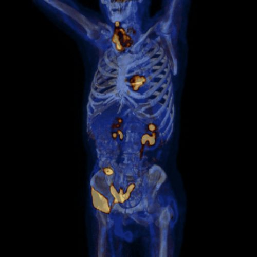
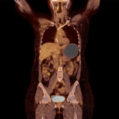
PET CT scan process - Full Body Positron Emission Tomography in Oncology
PET scans detect where FDG has accumulated in the body. Therefore, the PET tomography detects the location of metastatic cancer.

• Metastatic cancer is made of hypermetabolic cells.
• Hypermetabolic cells use more sugar (glucose) than normal cells.

• Radioactive glucose (FDG) is injected intravenously.

• During this resting period FDG collects in hypermetabolic cells.

• Table moves patient thru CT scanner ( 5 min.)
• Table moves patient thru FDG DETECTOR (15 min.)
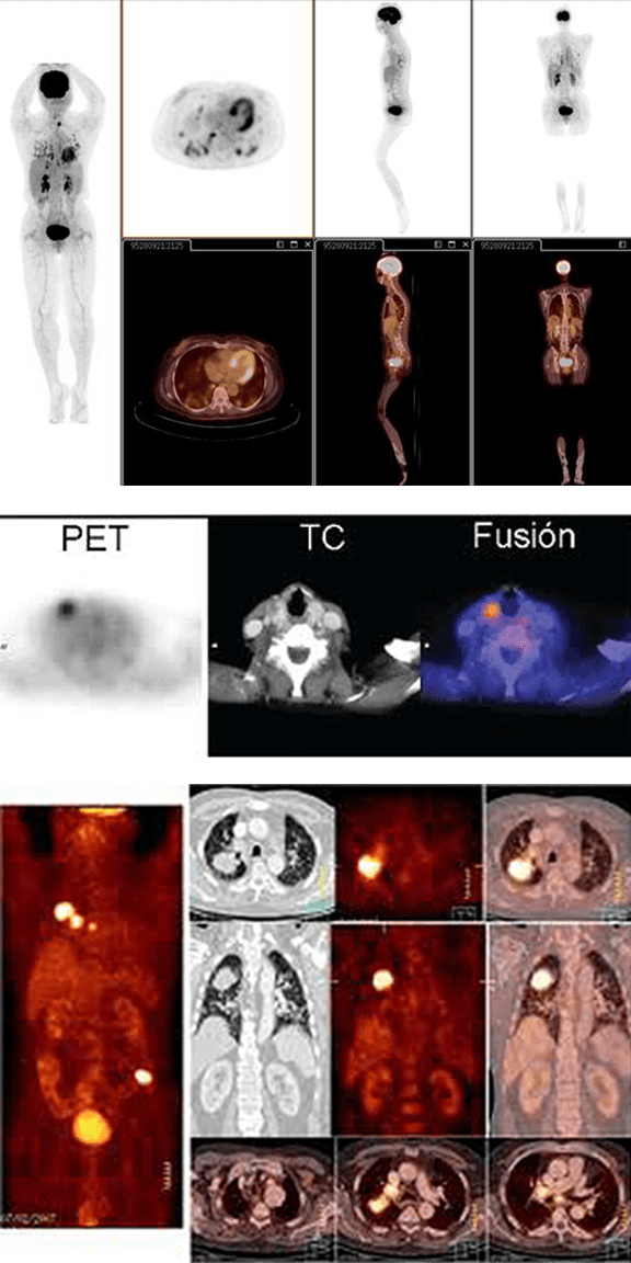
Examples of PET-CT images showing locations of organs and collected FDG:
Learn more about the Schedule Process for Full Body PET study
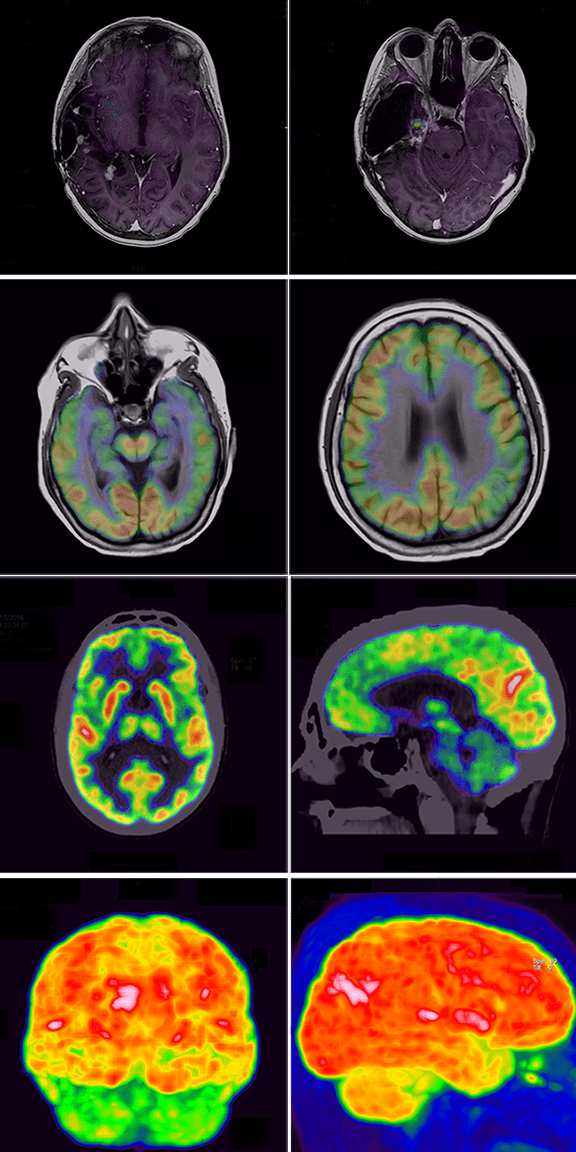
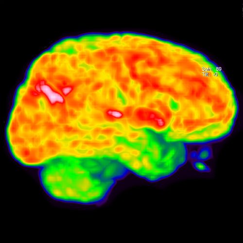
BRAIN PET scan process – Brain Positron Emission Tomography
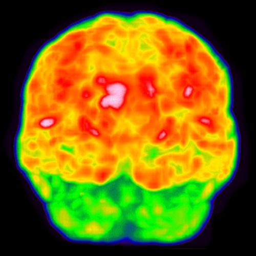
BRAIN PET tomography detects the distribution of FDG in the brain, locating areas of decrease or increase in brain metabolic activity in a precise and reliable manner.

• Brain metabolism consumes 25% of the total glucose that the body consumes. FDG or radioactive glucose, allows the detection of normal and abnormal metabolic areas in the brain.

• The radioactive glucose (FDG) is injected intravenously.

• During the relaxation period the radioactive glucose is distributed in the brain cells.

• Table moves patient thru CT scanner (2 min).
• Table moves patient thru FDG DETECTOR (for approximately 10 min).
Examples of BRAIN PET tomography images showing brain metabolism: